Microscope Magic
Today we looked at plant cells under a computer microscope. We found that best results were from red onions because we could clearly see the different cells.
After looking at the photographs, we observed that the cells were very regular in size (either a squashy rectangle or oval) believe that this is because of the rigid cell walls that plant cells have.
 |
| magnified x150 |
 |
| magnified x100 |

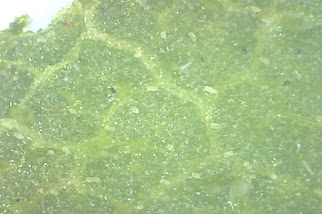
Top of a sunflower leaf Underside of a sunflower leaf
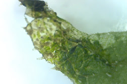 At the top left of this leaf are chloroplasts (the little green sacs)
Although we used a computer microscope we learnt some theory about light microscopes. This is:
😈Haha I have set homework for the home ed child!!! Hehe evil mum face (Please can you do for Mon 5/10/20?)
- A Specimen= The object your're putting under the microscope
- The Diaphragm= Controls the amount of light reaching the specimen
- The Objective Lens= To collect light from the sample
- The Eyepiece Lens=What you look through to see the specimen
- The Stageclips= They keeps the slides still
- Slide and Cover slide= Slides hold the specimen still you place one over the top of the other that has the specimen on it. That slide is called the cover slide
We also learnt a key practical skill-ALWAYS start on the lowest magnification. This is because it allows you to see more of the specimen then you can move the specimen around until you find the bit of it you want to zoom in on.
If you start at the strongest magnification then you are only seeing a very small part of the specimen and it is harder to locate the part of the specimen you are interested in studying.


Brilliant homework Jules! Completed on time and all right. XX The objective lens is tricky -I always remember it as the one which is closest to the object you are looking at (the specimen) contrasting with the eyepiece lens which is closes to your eye. Your explanation is better though xxx
ReplyDelete